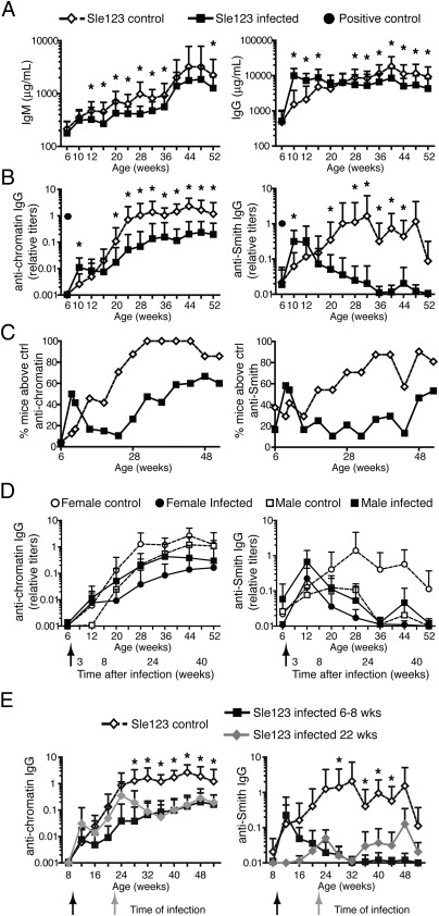
Fig. 3.
Chronic γHV68 infection significantly decreases autoantibody production in lupus-prone B6.Sle123 female mice. (A–D) Six- to 8-wk-old B6.Sle123 male (n = 5 per group) and female (n = 16–19 per group) mice were infected i.p. with 106 pfu of γHV68 or left uninfected. Blood was collected before infection, at 2 to 3 wk postinfection, and monthly for 11 mo from both groups of mice, and antibodies were analyzed by ELISA. (A) Total IgM and IgG in infected (filled square) and noninfected (empty diamond) B6.Sle123 female mice. (B) Anti-chromatin and anti-Smith IgG titers in serum of infected (filled square) and noninfected (empty diamond) B6.Sle123 female mice. A filled circle indicates the titer of anti-chromatin or anti-Smith antibodies in serum of a 15-wk-old female MRL/lpr (positive control). Data represent the mean and SD of antibody titers from 21 to 24 mice per group analyzed over the course of at least two separate experiments (*P < 0.05). (C) Frequency of B6.Sle123 female mice displaying anti-chromatin or anti-Smith antibody titers above mean titers + 2 SDs of B6 naive (i.e., control) mice. (D) Mean and SD of anti-chromatin and anti-Smith IgG titers in serum of infected (filled symbol) and noninfected (empty symbol) B6.Sle123 male (squares; n = 5 per group) and female (circles; n = 16–19 per group) mice. Arrows indicate the time of infection. (E) B6.Sle123 females were infected i.p. with 106 pfu of γHV68 at 8 wk or 5 to 6 mo (22 wk) of age. A group of females were left uninfected. Blood was collected starting at 6 to 8 wk of age and once per month for the time indicated. Shown are anti-chromatin and anti-Smith IgG titers in serum of B6.Sle123 females infected at 6 to 8 wk (filled black squares), at 22 wk (filled gray diamonds), and not infected (empty diamonds). Graphs represent the mean and SD of antibody titers from 10 to 20 mice per group analyzed in one to two experiments (*P < 0.05 between control and infected at 22 wk).

