Fig. 5.
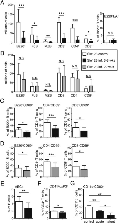
Chronic γHV68 infection decreases lymphocyte and DC activation in B6.Sle123 mice. (A–F) B6.Sle123 mice described in Fig. 3 that were infected at 6 to 8 wk or at 22 wk of age or left noninfected were euthanized at approximately 12 to 13 mo of age, at which time spleen cells were analyzed by flow cytometry as described in Fig. 1 and shown in Fig. S2B. (A and B) Absolute cell numbers of B- and T-cell subsets in B6.Sle123 female mice that were noninfected (A and B, white bars) or infected at 6 to 8 wk (A, black bars) or 22 wks (B, gray bars) of age. (A) Right: Percentage of Igλ+ B cells in the B220+ cell population of noninfected B6.Sle123 mice relative to mice that were infected at 6 to 8 wk of age. FoB, follicular B cells; MZB, marginal zone B cells. (C and D) Frequency of activated (CD69+) B cells (B220+), CD4, and CD8 T cells of B6.Sle123 female mice infected at 6 to 8 wk (C, black bars) or 22 wk (D, gray bars) of age relative to noninfected mice (white bars; n = 10–20 mice per group from separate experiments). (E) Spleen cells from B6.Sle123 female mice that were not infected (white bar) or infected at 6 to 8 wk of age (black bar) were analyzed by flow cytometry for the detection of ABCs (n = 10–16 mice per group from two separate experiments). ABCs were gated as single live and B220+CD4/CD8−CD11c+ lymphoid cells to determine their frequency. The bar graphs represent the arithmetic mean and SD of the percentage of ABCs in the B220+ B-cell population. (F) Frequency of FoxP3+ Tregs in the CD4+ T-cell population of the spleen of B6.Sle123 mice that were left noninfected (white bar) or infected at 6 to 8 wk of age (black bar; n = 14–24 mice per group from two separate experiments). (G) CD11c+ cells were purified from B6.Sle123 female mice and analyzed by flow cytometry for the expression of CD80 on CD11c+ cells. The graph represents the frequency of CD80+ cells in the total CD11c+B220− cells in the spleen of noninfected (control, white bar) mice and during acute (9 d postinfection) and latent (12 mo postinfection) γHV68 infection (n = 3 mice per group). An additional independent experiment performed on similar groups of mice (n = 3–5) resulted in the following frequencies of CD80+ cells in the CD11c+B220− cell fraction: control, 32.4%; acute, 19.5% (P = 0.01 vs. control); and latent, 11.8% (P = 0.0009 vs. control). Bar graphs represent arithmetic means and SDs (*P < 0.05, **P < 0.01, and ***P < 0.001; NS, not significant).

