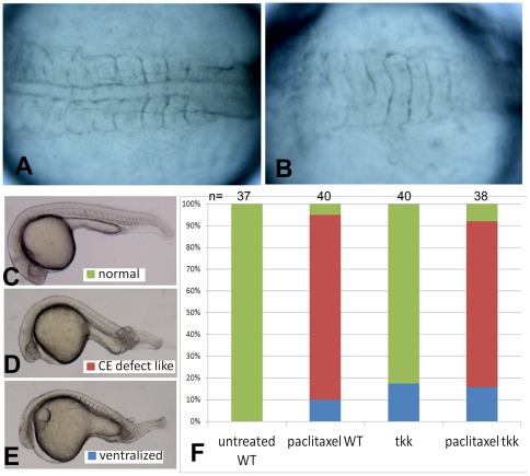
Figure 6. Paclitaxel treatment on wild-type and tkk mutant embryos.
(A) Dorsal view of an untreated 12 hpf embryo. (B) Dorsal view of a 0-mpf paclitaxel treated embryo with ventralized phenotype. (C) An embryo with normal phenotype. (D) An embryo with CE defect like phenotype, showing a smaller head, shorter anterior-posterior axis and malformed somites. (E) A ventralized embryo. Embryos in (C-E) were observed at 24 hpf. The statistical data based on the 24 hpf observation were shown in (F) with embryo numbers shown on the top of each column.

