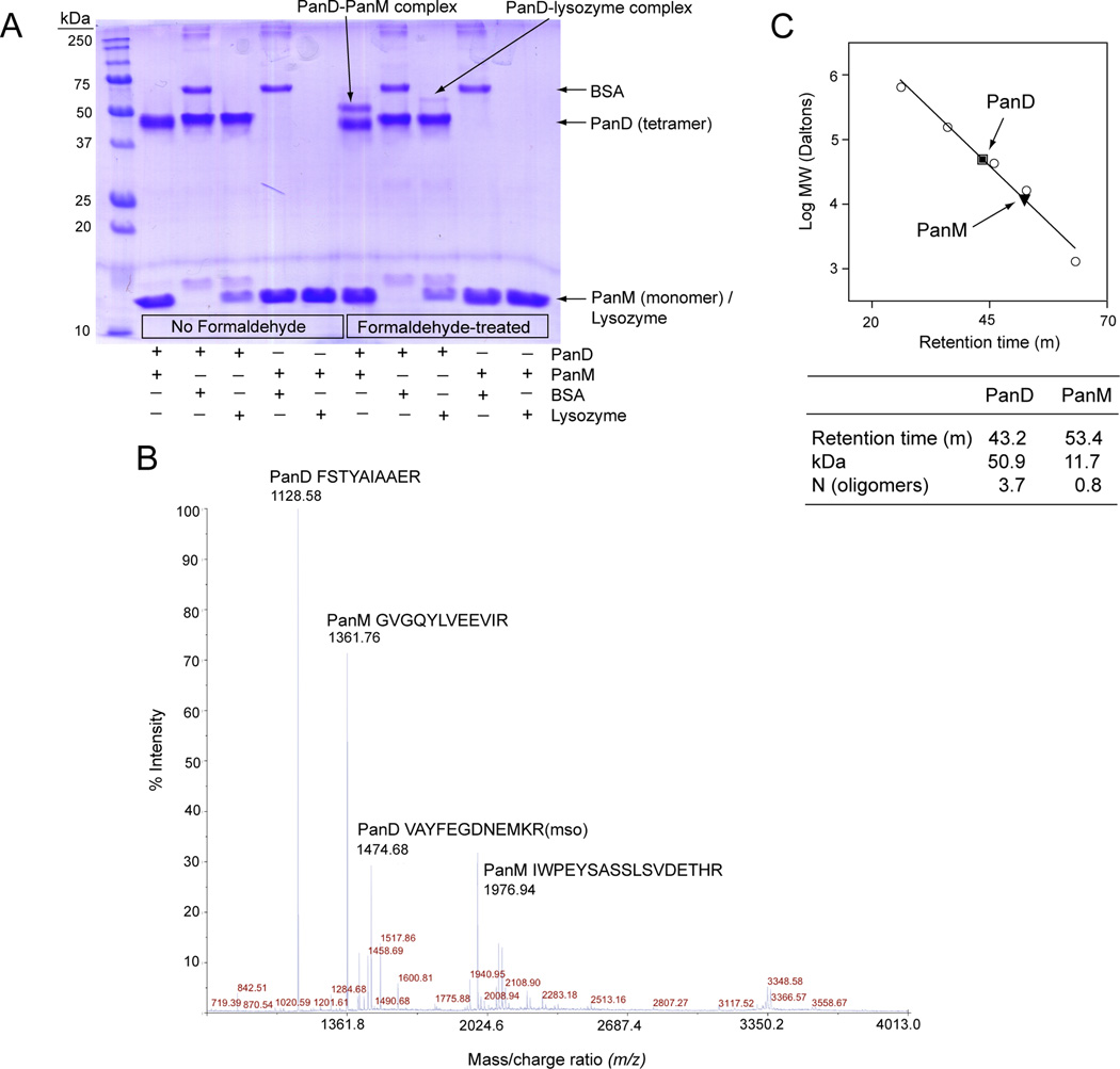
Figure 5. Interaction between PanD and PanM.
A. SDS-PAGE of formaldehyde crosslinking reactions. Lane 1 contained the Precision Plus Protein™ All Blue Standards (Bio-Rad). Lanes 2–6 contained untreated proteins, while lanes 7–11 contained formaldehyde-treated proteins (2 and 7: PanD+PanM, 3 and 8: PanD + BSA, 4 and 9: PanD + lysozyme, 5 and 10: PanM + BSA, 6 and 11: PanM + lysozyme). PanD appears as a tetramer in the denaturing gel because the samples could not be heated prior to running gel, which is necessary for tetramer denaturation. B. MALDI-TOF-TOF mass spectrum of the PanD-PanM complex shown in panel A. The amino acid composition and source of the four most abundant ions are indicated. Mso, methionine sulfoxide. C. Gel filtration analysis of PanD (square) and PanM (triangle). Molecular mass standards (circles) are thyroglobulin (bovine; 670 kDa), γ-globulin (bovine; 158 kDa), ovalbumin (chicken; 44 kDa), myoglobin (horse; 17 kDa) and vitamin B12 (1.35 kDa).

