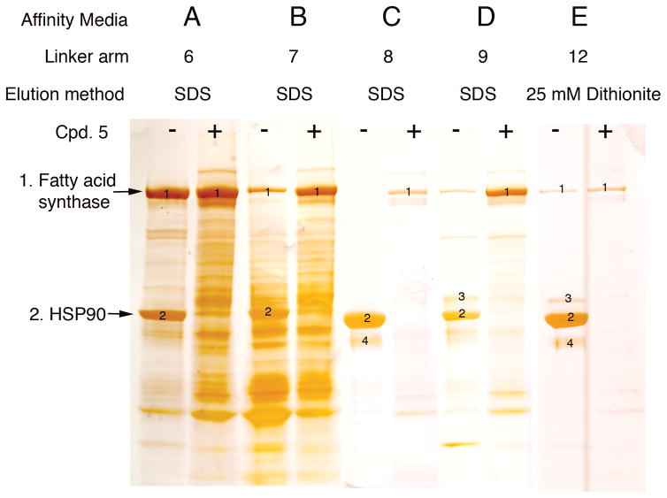
Figure 2. Directed chemical evolution of a selective affinity resin for Hsp90.
SDS-PAGE silver stain showing the effects of different side chain modifications on Hsp90 recovery and recovery of non-specifically bound proteins. Selectivity towards Hsp90 was demonstrated by inclusion of 1 mM 5 (+) in the tissue extract prior to mixing with affinity resin. Mass spectrometry was used to identify the bound proteins. In lane E bound proteins were eluted with 25 mM sodium dithionite in phosphate buffered saline. Numbers indicate bands that were sequenced by MS; (1) Fatty acid synthase (2) Hsp90 (3) Hsp90 (4) Hsp90 proteolytic fragments.

