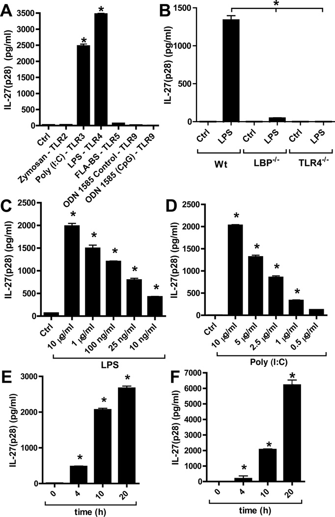
Figure 1.
Characterization of IL-27(p28) release from macrophages after activation of TLR3 or TLR4. (A) Peritoneal elicited macrophages (PEM) from C57BL/6J (Wt) mice were incubated with agonists for various TLR-agonists (all 1 µg/ml) and secreted IL-27(p28) detected by ELISA after 10h. (B) Macrophages from Wt mice, LBP−/− mice and TLR4−/− mice were incubated with LPS (50ng/ml) for 10 h before detection of IL-27(p28). (C) Dose response studies of IL-27(p28) release by PEM after TLR4-activation by LPS (10 µg/ml – 10 ng/ml), 10 h. (D) Dose response curve of IL-27(p28) production after TLR3-activation by Poly (I:C) (10 µg/ml – 500 ng/ml), 10 h. (E) Time course of IL-27(p28) release after LPS (1 µg/ml). (F) Time course of IL-27(p28) production after Poly (I:C) (10 µg/ml). All experiments shown were done with thioglycollate elicited PEM from C57BL/6J (Wt) mice. Data are representative of at least 3 independent experiments. *p < 0.05, error bars represent s.e.m.

