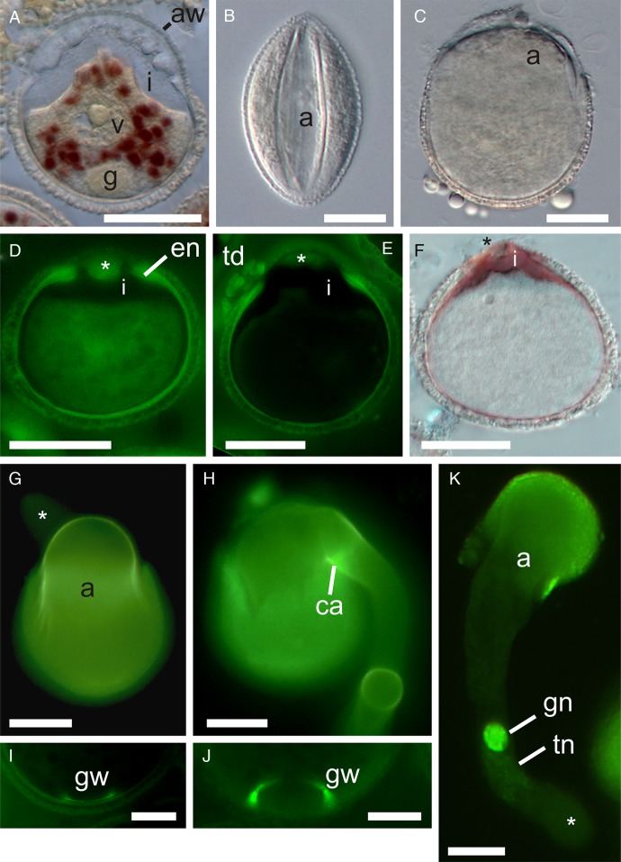
Fig. 2.
Pollen germination of A. scandens. (A) Two-celled pollen from female phase flower (closed anther) with IKI-stained granules in vegetative (tube) cell cytoplasm. (B) Pollen from open anther in immersion oil, showing dehydrated state at presentation (DIC). (C) In vitro hydrated pollen, just before germination (DIC). (D) Immature pollen (female phase of flower) with AB staining of extra-apertural endexine (en) and isolated endexine in aperture wall (asterisk). (E) Mature pollen from open anther (AB). Note AB stain in a thin layer of endexine which is thickened at the aperture edge, absent in its margins and present in the centre of the apertural wall (asterisk). Clumps of AB-stained material (of tapetal origin) are associated with the outer apertural wall. Note also the bulging of intine through aperture, probably caused by partial hydration of pollen during fixation. (F) Pollen from open anther showing intine stained by ruthenium red. (G) In vitro germinated pollen showing AB stain in inner pollen wall and its continuity with the emerging inner tube wall. Note that the tube tip (asterisk) in the background lacks AB staining. (H) Emergence of pollen tube has ruptured part of pollen wall and pushed aside aperture covering. Note strong AB staining of a presumably callose annulus (ca) at base of tube. (I, J) A ring of AB-stained material is prominent at the proximal pole of many mature and germinated pollen grains, marking the location of the generative cell wall (gw). Scale bars = 10 µm. (I) Callose wall of generative cell inside of intine. (J) Remnants of callose wall of generative cell. (K) In vivo germinated pollen (3 hap) with faintly stained tube nucleus (tn) in association with the generative cell near the young pollen tube tip (asterisk) (DAPI). Pollen from (G), (H), (J) and (K) was fixed and stained 2 h after innoculation on growth medium. (A), (D–F) and (I) are from methacrylate sections. a, aperture; aw, aperture wall; g, generative cell; gn, generative cell nucleus; i, intine; td, tapetal deposits; v, vegetative cell nucleus. Scale bars = 20 µm, except where noted.

