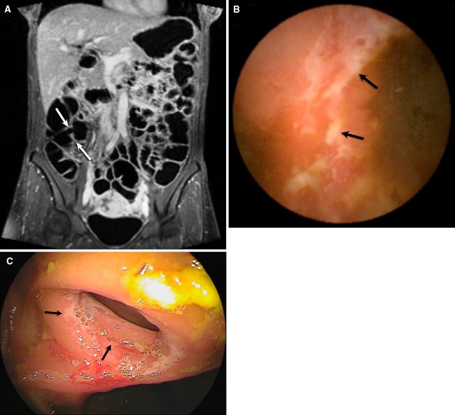
Fig. 3.
39-Year-old female patient with known CD and postoperative ileocecal resection 3 years earlier. Patient complaints were abdominal pain and diarrhea. MRE showed on coronal T1 3d fat-sat image (A) after contrast injection, normal anastomosis (arrows) of the neo-ileocecal junction without bowel wall thickening or increased contrast enhancement. CE (B) and BAE (C) both indicated superficial ulcerations on the level of the anastomosis (arrows). No other abnormalities were diagnosed.

