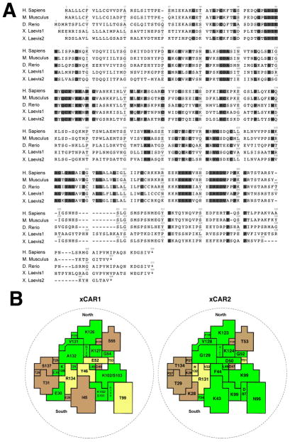
Figure 1. Sequence analysis.
A. Predicted amino acid sequences of the Xenopus laevis Coxsackievirus and Adenovirus Receptors (xCAR1 and 2). The xCAR sequences are shown aligned with CAR proteins from mouse, human and zebrafish. The shaded residues are identical in all these sequences.
B. Diagram of the binding site for virus on the CAR surface, based on the structure of hCAR in (He et al., 2001). The numbering is for the Xenopus sequences and the color depicts the relatedness to the human sequence: bright green identical, yellow conserved, brown unrelated. (structural biology method adapted from (Petrella et al., 2002)).

