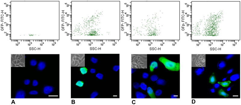
Figure 2. Evidence that xCARs function as adenovirus receptors.
The lower panels show GFP fluorescence in the cells, the upper show flow cytometry of the above-threshold cells by GFP intensity (vertical axis) and cell size (horizontal axis). The number of GFP positive cells in the presence of xCAR is similar to the transfection efficiency (about 5%), indicating that all or most cells transfected with xCAR become infected. All cell nuclei are stained blue with DAPI.
A. CHO cells treated with lipofectamine alone and infected with adeno-GFP (negative control).
B. CHO cells transfected with pcDNA3-GFP (transfection control).
C. CHO cells transfected with xCAR1, and then infected with adeno-GFP.
D. CHO cells transfected with xCAR2, and then infected with adeno-GFP. Scale bars 5 μm.

