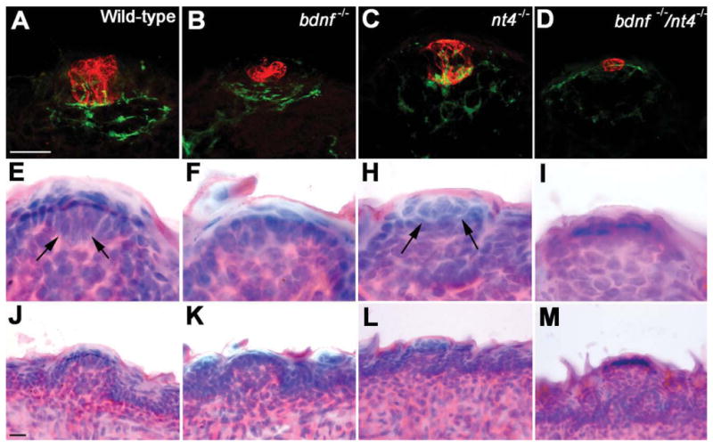
Figure 4.

The morphology of the remaining taste buds is more sensitive to deletion of BDNF than NT-4. Images of taste buds in the tongue fungiform papillae in mice at birth were visualized using double immunolabeling for anti-cytokeratin-8 (developing taste bud; red) and anti-GAP-43 (innervation; green) (A-D). Compared to wild-type taste buds, the developing taste buds in Bdnf−/− (B) and Bdnf−/−/Ntf4/5−/− (D) mice show a drastic reduction in overall size. The taste buds in Ntf4/5−/− (C) mice are normal (similar to the wild-type taste buds in A in appearance). The morphology of some taste buds was visualized using H&E staining (E–I). Note that the taste buds in wild-type (E) and Ntf4/5−/− (H) mice are recognizable based on their morphology in the H&E-stained images (arrows). However, anti-cyto-keratin-8-positive clusters in Bdnf−/− (B) and Bdnf−/−/Ntf4/5−/− (D) mice do not morphologically resemble taste buds in H&E-stained tissue (F,I). All taste buds are located in clear fungiform papillae (J-M). A magenta-green copy of this image is provided as Supporting Figure 1. Scale bars = 10 μm in A,J.
