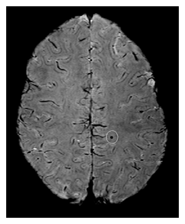
Figure 1.

Hemosiderin detected with multiecho susceptibility weighted imaging using a 3 Tesla scanner. Multiecho SWI image (Philips Achieva 3T; 5 echoes; voxel size = 0.32 × 0.32 × 0.75 mm3). Courtesy of Alexander Rauscher, Ph.D., UBC MRI Research Centre, Department of Radiology, University of British Columbia, Vancouver, Canada.
