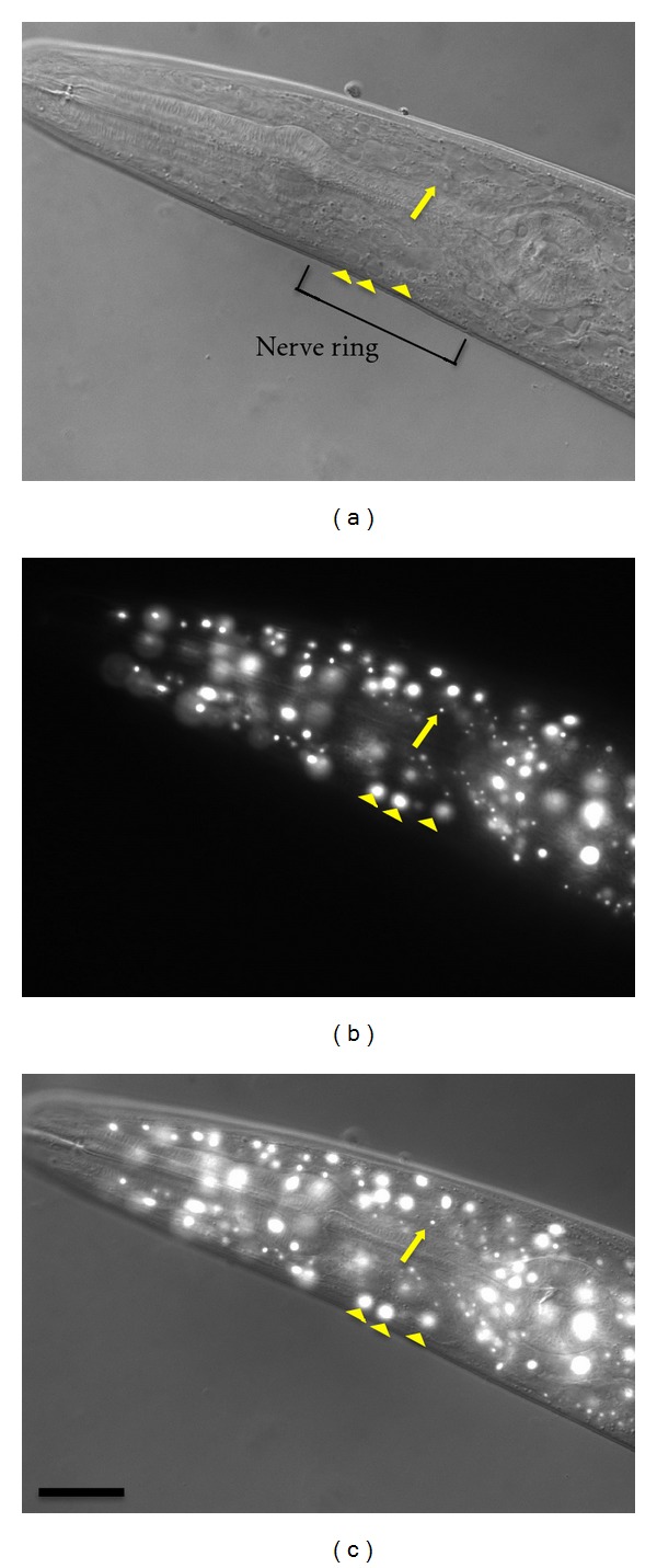
Figure 2.

Comparison of nucleolar appearance of cells in the head region in Nomarski and fluorescence micrographs. A transgenic worm expressing nucleolar protein FIB-1::GFP was photographed using Nomarski and fluorescent microscopy. An arrow indicates the nucleolus of neuronal cells located at the nerve ring (as indicated) while arrowheads indicate the nucleoli of the hypodermis in (a) Nomarski micrograph, (b) fluorescence micrograph, and (c) merged (a, b) images. The scale bar indicates 20 μm.
