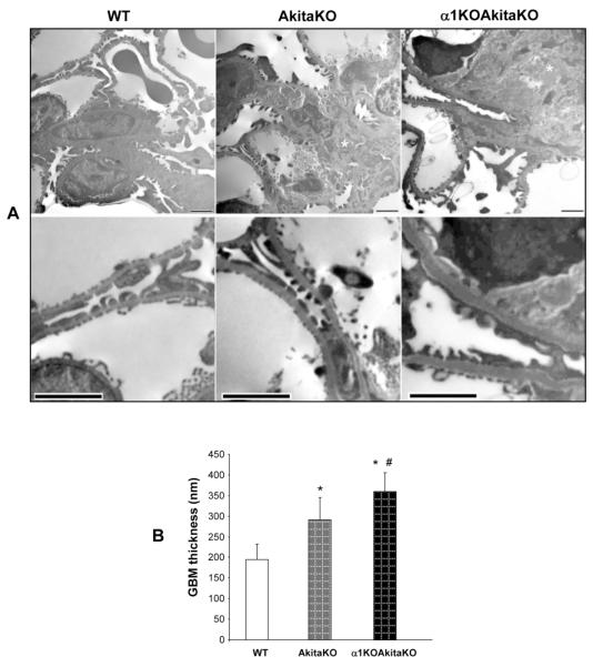
Figure 6. Increased mesangial matrix expansion and GBM thickening in α1KOAkitaKO Balb/c mice.
(A) Electron microscopy pictures of glomeruli of 6 month old WT, AkitaKO and α1KOAkitaKO mice. The upper panels demonstrate increased mesangial expansion (asterisk) in the AkitaKO mice, which is significantly worse in the α1KOAkitaKO mice. Scale bar, 2 μm. Obvious increase in the thickness of the GBM was evident in both AkitaKO and α1KOAkitaKO mice at 6 months (lower panel in A; scale bar, 2 μm) although it was more prominent in the latter group (B). Values in B represent the mean +/− SD of 30 measurements. Differences between (*) or AkitaKO vs. α1KOAkitaKO mice (#) were significant (p<0.05).

