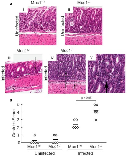
Figure 1.
H. pylori-infected Muc1−/− mice have increased neutrophils in the gastric mucosa compared with Muc1+/+ mice. (A) Muc1+/+ (i,iii) and Muc1−/− (ii,iv) mice were uninfected (i,ii) or were infected with H. pylori (iii,iv), and gastric mucosa were assessed at 4 weeks post-infection by Giemsa staining. Arrows indicate inflammatory cell infiltrates. Original magnification, 200×. (v) Illustrates a higher magnification of the indicated area of (iv) to demonstrate the infiltrating cells. (B) Quantitative analysis of gastric inflammation on a scale of 0–5 based on the following parameters: 0, no significant lesions; 1, mild infiltrate of inflammatory cells; 2, larger focus of inflammation extending between glands and/or in submucosa; 3, patches of inflammation extending between glands toward the lumen and in the underlying submucosa; 4, intense transmucosal inflammatory infiltrate extending across the field distorting glandular architecture; 5, extensive mucosal and submucosal inflammation with disruption of glandular architecture and ulceration (Stuller et al., 2008). Each symbol represents an individual tissue section. Horizontal bars represent median values. Differences between median values were assessed by the Student’s t test. The results are representative of two experiments.

