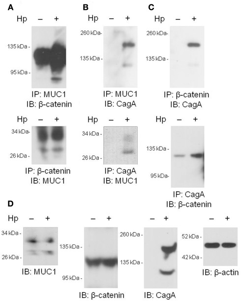
Figure 2.
Co-immunoprecipitation of MUC1, β-catenin, and H. pylori CagA. (A–C) AGS cells were untreated or were treated with H. pylori (Hp) at an MOI of 100. At 24 h post-infection, the cells were washed and cell lysates were immunoprecipitated with the indicated antibodies. Immunoprecipitated proteins were resolved by SDS-PAGE, transferred to PVDF membranes, and processed for immunoblotting with the indicated antibodies. (D) Lysates of untreated or HP-treated AGS cells were resolved by SDS-PAGE, transferred to PVDF membranes, and processed for immunoblotting with antibodies to MUC1, β-catenin, CagA, or β-actin. Molecular weight markers are indicated on the left in kilodaltons (kDa). The results are representative of two experiments.

