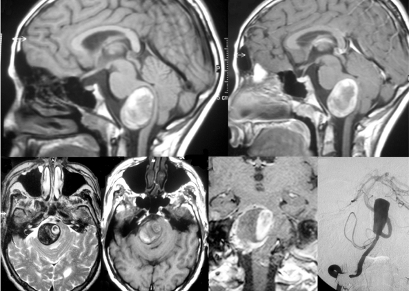
Figure 2.
This patient became symptomatic due to brain stem compression, MRI (T1 pre contrast enhancement in frames A and D, post-contrast in frames B and E and T2-weighted images in frame C) shows the typical findings of a partially thrombosed aneurysm of the V4 segment. Methemoglobin (T1 hyperintense on pre-contrast images) as a sign for an acute bleeding is present at the rim of the aneurysm far from the perfused part that is in this case located medially. T2-weighted images show perifocal edema and the onion-skin layer of mural thrombus of different ages.

