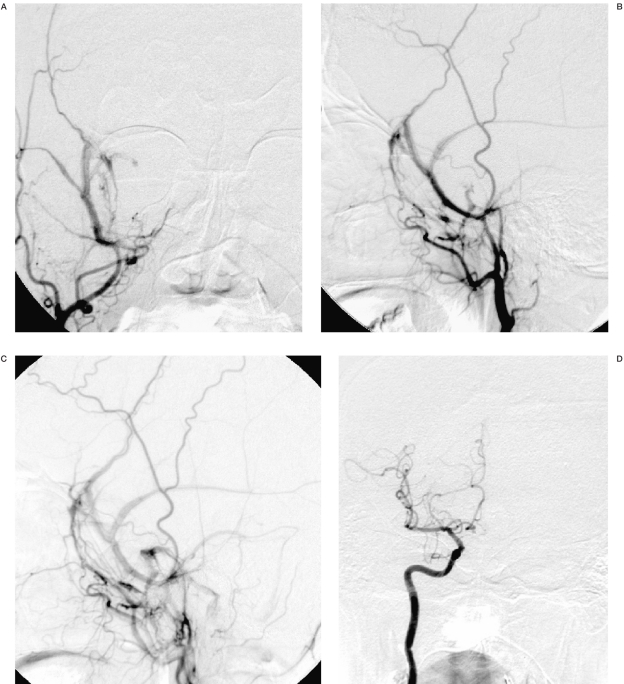
Figure 2.
Right external carotid artery angiogram AP (A), lateral (B) shows middle meningeal arteriovenous fistula with drainage towards sinus of lesser wing of sphenoid, cavernous plexus, pterygoid plexus and visualization of petrosal and meningeal veins in later phase (C). Right Internal Carotid angiogram (D) is normal.

