
Figure 3.
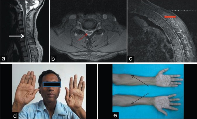
Patient no. 8 – A 50-year-old male with weakness and wasting both hands and forearms for 2 years – neutral position sagittal T2WI magnetic resonance imaging (MRI) showing loss of cervical lordosis and localised lower cervical cord atrophy (white arrow) (a), axial T2WI MRI showing anteroposterior cord flattening with loss of attachment of dura from subjacent lamina (thin red arrow) (b), flexion contrast MRI showing posterior epidural thin crescentic enhancing region (thick red arrow) (c), wasting of both hands (d) and forearms with oblique amyotrophy (black arrows) (e)