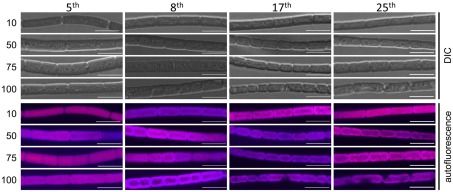
Figure 2.
Confocal laser scanning microscopy analyses of cellular morphology of Fremyella diplosiphon UTEX 481 wild-type (WT) strain under green light (GL) conditions of varying light intensity. F. diplosiphon WT was grown in BG-11 culture medium containing 20 mM HEPES at 10, 50, 75, or 100 μmol m−2 s−1 (numbers indicated at left) at 28°C with shaking at ∼175 rpm as indicated in Figure 1. Representative optical slices from a Z-series of differential interference contrast (DIC) images (upper half) and maximum intensity projection of phycobiliprotein autofluorescence images (lower half) of WT. Numbers of consecutive dilutions (5, 8, 17, or 25) made before imaging are indicated at the top of each column. All images were acquired with a 40× oil objective with 2× zoom. Bar represents 10 μm.

