Figure 3.
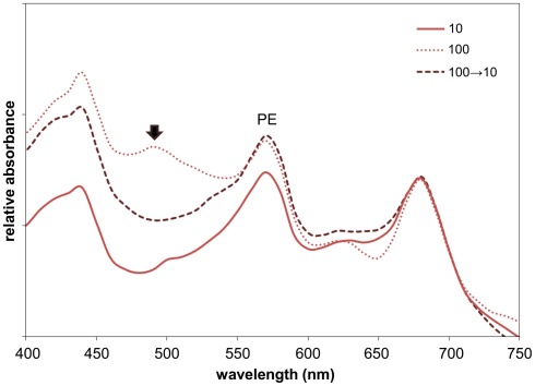
Whole-cell absorbance spectral scans of Fremyella diplosiphon UTEX 481 wild-type (WT) strain under green light (GL) conditions of varying light intensity. Representative whole-cell spectral scans of WT cells grown at 28°C with shaking at ∼175 rpm in GL at 10, 100 μmol m−2 s−1, or at 100 μmol m−2 s−1 then moved to 10 μmol m−2 s−1 for 3 days (100 → 10). Absorption maxima for phycoerythrin (PE, ∼565 nm) is indicated. The left and right-most peaks (∼430 and 680 nm, respectively) are chlorophyll absorption peaks. The black arrow indicates the peak at ∼480–490 nm that appears in cells under high GL intensity.

