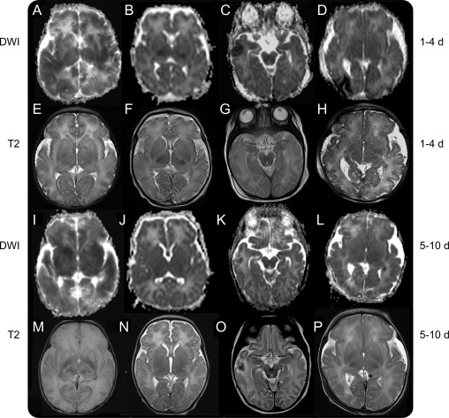
Figure 1. Images from 4 patients scanned at 2 timepoints.
The first presented with global injury (A, E, I, and M); the second presented with basal ganglia and posterior limb internal capsule (PLIC) injuries (B, F, J, and N); the third presented with cortical injury (C, G, K, and O); the fourth presented with border zone injury (D, H, L, and P). Diffusion imaging during the first 4 days after birth (A–D) shows respectively global (cortex and deep nuclear gray matter), bilateral PLIC, right temporal cortex, and right occipital border zone reductions in mean diffusivity (MD) with resolving reductions in MD seen in the delayed images obtained at 5–10 days after birth (I–L). The most persistent reduction in MD is seen in the global injury case (I). Corresponding T2-weighted images during the first 4 days after birth (E–H) showed mild lesions, more conspicuous in T2 images obtained after day 4 (M–P), demonstrating respectively a complete loss of cortical ribbon and an absence of differentiation between white and gray matter, absence of normal ventral thalami nuclei and PLIC signal (M), an absence of normal signal in the PLIC (N), a hypointensity surrounded by hyperintensity in right temporal lobe suggestive of hemorrhagic infarct (O), and subtle signal abnormality in the right occipital region (P). DWI = diffusion-weighted imaging.

