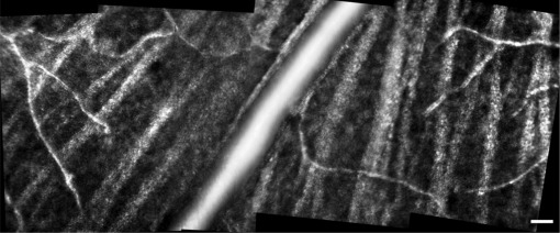
Fig. 6.
In vivo reflectance image montage of the NFL in the mouse eye, showing a large blood vessel in the center, capillaries, and nerve fiber bundles. This location was over 15 degrees away from the optic disk. Size of this image was 553 µm × 230 µm, or 16.3° × 6.8°. Each individual image was an average of 50 frames. Confocal pinhole diameter was 2.1 Airy disks. Scale bar: 20 µm. Image was contrast stretched for display purposes only.

