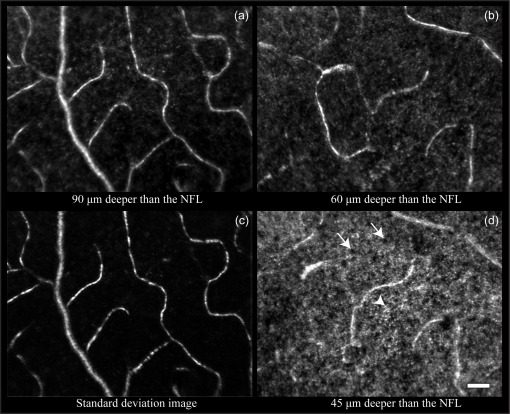
Fig. 7.
In vivo reflectance capillary images in the mouse retina. All images are taken at the same retinal location. (a), (b), (d) Capillary images at different depths. Each image is a registered average of 50 individual frames. (c) Standard deviation/motion contrast image corresponding to the depth of image (a). Arrows and arrowhead: dark regions and microscopic bright point structures within the intermediate capillary layer. Confocal pinhole diameter was 2.1 Airy disks. All images were contrast stretched identically for display purposes. Scale bar: 20 µm.

