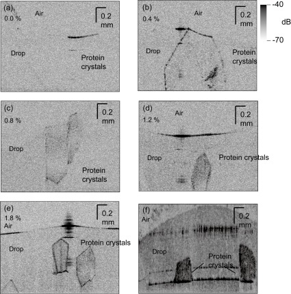
Fig. 2.
Cross sections of three-dimensional UHR-OCT images of HEWL crystals grown in (a) 0.0, (b) 0.4, (c) 0.8, (d) 1.2, and (e) 1.8% (w/v) agarose. (f) An image of a drop that contained Synechococcus phosphoribulokinase crystals and 1.0% (w/v) agarose. The crystals shown in panels (e) and (f) are clearly and non-invasively seen at μm resolution. Media 1 (3.7MB, MOV) shows the 3D UHR-OCT image of HEWL crystals grown in the same condition as that for Fig. 2(e).

