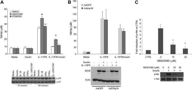
Fig. 3.
Role of p38 and MAPK p42/p44 in the regulation of iNOS by insulin. A: hepatocytes were pre-incubated with MAPK p38 inhibitor SB203580 (10 μM) or MAPK p42/p44 inhibitor PD98059 (10 μM) for 30 min and then cultured with IL-1β + IFN and insulin (10 μM) for 24 h. The supernatants were collected for nitrite (top). Bottom: hepatocytes were treated with TNF-α (500 U/ml), IL-1β (200 U/ml), IFN (100 U/ml), or insulin (10 μM) for 30 and 60 min, then analyzed by Western blot for phosphorylated and total p38. *Significant difference vs. DMSO (P < 0.05). B: hepatocytes were infected with AdGFP (light gray) or AdDN-p38 (dark gray) with MOI (10:1) for 24 h. The cells were then treated with IL-1β + IFN with or without insulin (10 μM) for another 24 h. The supernatants were collected for nitrite, and cellular proteins were collected for Western blot. C: hepatocytes were cultured with insulin (10 μM) and SB203580 at the indicated concentration for 30 min. Cellular proteins were then collected for Western blot and quantitated by densitometry.

