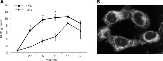
Fig. 1.
A: curcumin uptake (50 μM, 0–30 min) was performed with T-84 cells at 37°C or 4°C. Similar results were obtained in conditionally immortalized mouse colonocytes [young adult mouse colonocytes (YAMC); not shown]. Cells treated with DMSO for respective durations were used for calculating background fluorescence. *Statistically significant differences (P ≤ 0.05) between curcumin uptake at 37°C and 4°C at respective time points (Student's t-test; n = 6). B: z-section from live cell imaging of curcumin-loaded YAMC cells 20 min into 50 μM curcumin treatment.

