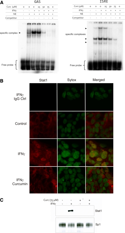
Fig. 5.
EMSA analysis of nuclear protein binding to the GAS and IFN-stimulated regulatory element (ISRE) cis-elements. A: cells were treated with IFN-γ for 30 min without or with increasing concentrations of curcumin (25–75 μM; administered 20 min prior to the addition of IFN-γ). NE, nuclear extract; Competitor, 100 × excess of unlabeled probe. Excess of labeled probe indicated at the bottom. Immunofluorescence (B) and Western blot (C) analysis of Stat1 nuclear translocation in T-84 cells. Cells were treated with curcumin (75 μM) and IFN-γ in a way analogous to that used in EMSA analysis (A). Green nuclear staining is Sytox green. Red staining represents Stat1 (Alexa647). Topmost panels show negative (rabbit IgG) control. Sp1 was used as a loading control for Western blot detection of total nuclear Stat1 (C).

