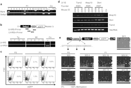
Figure 3.
No ectopic transgene transcription and expression in transgenic mice due to extensive methylation of spleen focus-forming virus promoter (SFFVp). (a) Genotyping-PCR analyzing tail-tip DNA of F1 mice. (b) Scheme of primer localization and reverse transcription-PCR (RT-PCR) of transgenic (Tg) and wild-type (Wt) F1 mouse whole body slices; Co, controls. (c) Peripheral blood of 49 mice was analyzed for enhanced green fluorescent protein (eGFP) expression (six representative fluorescence-activated cell sorting (FACS) blots are shown here). (d) RT-PCRs verifying transcription of genes adjacent to the proviral vector integration in progeny of three different transgenic founder mice (founder mice 323, 325, and 347). Mouse numbers in black represent transgenic mice, numbers in gray the wild-type counterparts. LV IS, lentiviral integration site. (e) Scheme of transgenic LV and the analyzed CpGs within promoter region. Heat maps for 454 reads (rows) show the methylation status of 18 CpGs (columns) within the SFFVp region in transduced control (SC1) cells and in transgenic animals (n = 8 representatives). Methylated CpGs are shown in dark gray, unmethylated CpGs in light gray and gaps in white. The average methylation level for each CpG site is visualized in the lower bar using a light gray to black color scale. FSC, forward scatter.

