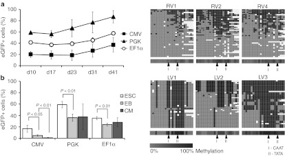
Figure 5.
Silencing of virally derived internal promoters during differentiation in lentivirally transduced embryonic and adult stem cells. (a) Enhanced green fluorescent protein (eGFP) expression over time in undifferentiated mouse embryonic stem cells (ESC) after transduction of CMV, PGK, or EF1α driven lentiviral vectors (MOI 50). (b) Percentage of eGFP+ cells during differentiation of transduced ESC with CMV, PGK, and EF1α driven lentiviral vectors. CM, cardiomyocytes; EB, embryoid bodies. (c) Heat maps for 454 reads (rows) show the methylation status of 26 CpGs (columns) within the spleen focus-forming virus promoter (SFFVp) in lentiviral vector (LV) and γ-retroviral vector (RV) backbone, 20 weeks after bone marrow transplantation. Methylated CpGs are shown in dark gray, unmethylated CpGs in light gray and gaps in white. The average methylation level for each CpG site is visualized in the lower bar using a light gray to black color scale.

