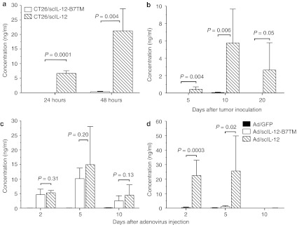
Figure 8.
Systematic and local expression of IL-12. (a) 5 × 10⁴ CT26/scIL-12-B7TM or CT26/scIL-12 cells were seeded on 6-well plate and the supernatants collected 24, or 48 hours later to measure the amount of IL-12 by ELISA. The values are presented as the mean ± SD of triplicate cultures. (b) Groups of BALB/c mice (n = 5) were injected subcutaneously with 1 × 10⁷ CT26/scIL-12-B7TM, or CT26/scIL-12 cells, then serum samples were collected on the indicated days and assayed for the presence of IL-12. (c,d) Mice (n = 3–6) with established subcutaneous CT26 tumors as described in Figure 5 were injected intratumorally with 1 × 10⁹ pfu of Ad/scIL-12-B7TM, Ad/scIL-12, or Ad/GFP on day 10. (c) Tumor and (d) serum samples were collected on the indicated days and assayed for the presence of IL-12 by ELISA. The results in b–d are the mean ± SD for the indicated number of mice. ELISA, enzyme-linked immunosorbent assay; IL, interleukin.

