Figure 2.
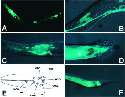
Expression of a BRA-1∷GFP Fusion. (A) BRA-1∷GFP expression of an L3 hermaphrodite. Strong expression was observed in the head region and the tail region. (B) Strong signals were seen in the ventral nerve cord. (C) Lateral view of the adult hermaphrodite showing GFP expression in the head. GFP fluorescence was observed in the cell bodies of BAG, AFD, ASK, ASI, ASG, ASE, ASJ, and AWC neurons. (D) Tail region of an adult male. GFP fluorescence was seen in unidentified male-specific neurons. (E) A diagram summarizing the BRA-1∷GFP-expressing neuron. (F) Tail region of adult hermaphrodite. GFP fluorescence was seen in the cell bodies of PHA and PHB phasmid neurons. Anterior is to the left, and dorsal is up.

