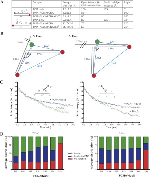
FIGURE 6.
Cleavage dynamics of PabNucS-PCNA complex on both 5′ flap and 3′ flap. A, summary of FRET measurements between 5′ and 3′ extremities of duplex DNA in both 5′ flap and 3′ flap substrates. B, FRET measurements define kinked DNA binding to the PabNucS-PCNA complex for both 5′ flap and 3′ flap. PabNucS binds upstream from the 5′ flap, whereas it binds downstream from the 3′ flap. C, cleavage activity of PabNucS in the presence (blue triangle) or absence (green triangle) of PabPCNA on both 3′ flap (right) and 3′ flap (left) detected by a decrease in anisotropy. D, cleavage activity of PabNucS on 5′ (right) and 3′ (left) flaps with and without PabPCNA. The reactions were performed using 1 μm PabNucS and the PabPCNA/PabNucS molar ratio was varied. The incubation time was 20 min. Cleavage products were analyzed and quantified using 18% denaturing polyacrylamide gels (9).

