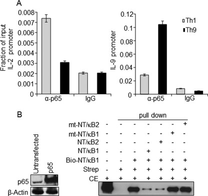
FIGURE 6.
In vivo and in vitro binding of NF-κB (p65) to the IL-9 promoter. A, ChIP assay was performed with in vitro differentiated and PMA/ionomycin-stimulated Th1 and Th9 cells using control IgG and NF-κB (p65) antibodies. The amounts of precipitated DNA were measured by qRT-PCR with primers specific for the IL-9 and IL-2 promoter regions. Relative NF-κB (p65) enrichment in the precipitated samples compared with total chromatin (input) is shown as a fraction of input. B, left, antibodies against NF-κB (p65) and actin (control) were used to perform Western blot for detecting NF-κB (p65) expression in untransfected and NF-κB (p65)-transfected HEK cells. Right, biotin-conjugated probe corresponding to NF-κB (p65) binding site 1 (NT/κB-1; −315/−306 in Fig. 1B) were incubated with NF-κB (p65)-overexpressing HEK-293 cell lysate in the absence or presence of the indicated non-biotinylated competitor probes. The protein-DNA complexes were precipitated with streptavidin (Strep) and analyzed by immunoblotting with α-NF-κB (p65) antibody. The first lane indicates input, which is 2.5% of the total cell extract (CE) used for pull-down. The data are representative of three independent experiments.

