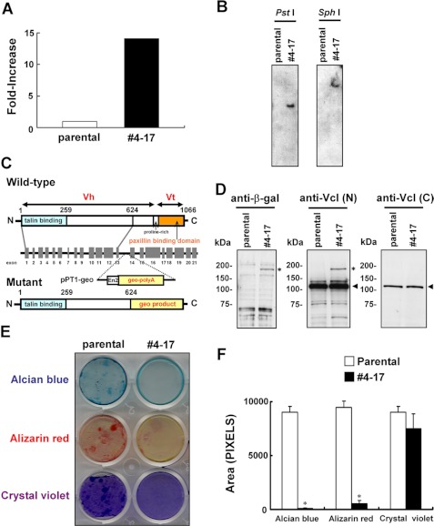
FIGURE 1.
Isolation of the clone with the trapped vinculin gene. A, the β-galactosidase activity in clone 4-17 and the parental ATDC5 cells. B, Southern blotting. Genomic DNA was digested with the restriction enzyme PstI or SphI, and subjected to hybridization with a radiolabeled fragment of lacZ cDNA prepared by EcoRI/SacI digestion of pPT1-geo. C, genomic organization of the murine vinculin gene and the insertional mutation resulting from the gene trapping. The locations of exons (gray boxes) and introns (horizontal lines between exons) are indicated. pPT1-geo was inserted into intron 13, resulting in a mutant protein in which vinculin lacking the tail domain was fused to the geo product. D, detection of the β-galactosidase fusion protein. Cell lysates were subjected to immunoblotting using antibodies against β-galactosidase (left) or amino-terminal (center) or carboxyl-terminal vinculin. The asterisk and the arrow indicate the signals corresponding to the fusion protein comprising part of vinculin and the geo product and that for wild-type vinculin, respectively. E, impaired chondrogenic nodule formation in clone 4-17. The cells were cultured for 8 weeks in chondrogenic medium and subjected to Alcian blue and Alizarin red staining. Clone 4-17 accumulated less cartilaginous matrix than parental cells. The cells were also stained with crystal violet to confirm the adhesion to culture plates. F, the stained area in E was quantitated using NIH Image 1.63 software. Data are shown as the mean ± S.D. (error bars) (n = 3). *, p < 0.001 versus parental cells.

