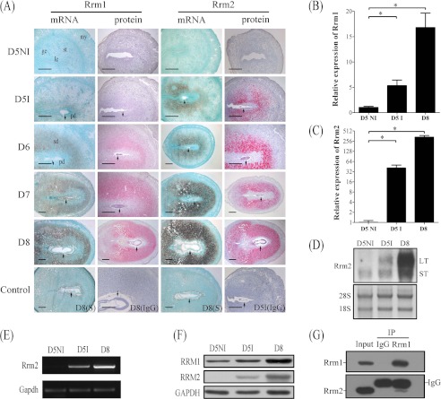
FIGURE 1.
Expression of RRM1 and RRM2 in mouse uterus during early pregnancy. A, both mRNA and protein localizations of RRM1 and RRM2 in mouse uterus from day 5 (D5) to day 8 (D8) were detected by in situ hybridization and immunostaining, respectively. Real-time RT-PCR was performed to quantify the mRNA level of Rrm1 (B) and Rrm2 (C) in mouse uterus on day 5 (D5NI, interimplantation site on day 5; D5I, implantation site on day 5) and day 8. D, Northern blot. Both long transcripts (LT) and small transcripts (ST) of Rrm2 were up-regulated at the implantation sites. 28S was used as internal reference. E, RT-PCR analysis of Rrm2 mRNA level in mouse pregnant uteri. Strong Rrm2 bands were observed in implantation sites on days 5 and 8 compared with the interimplantation site on day 5 of pregnancy. F, Western blot of RRM1 and RRM2 protein in mouse uterus on days 5 and 8. G, physical interaction between RRM1 and RRM2 in mouse decidua was detected by co-immunoprecipitation. ge, glandular epithelium; le, luminal epithelium; my, myometrium; st, stroma; pd, primary decidua; sd, secondary decidua; arrow, embryo. Bar, 300 μm. *, p < 0.05; error bars, S.E.

