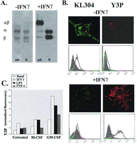
Figure 3.
Expression and conformation of class II MHC molecules are regulated by proinflammatory cytokines. (A) IFN-γ induces expression of SDS-stable MHC class II on N9 cells. Cells were incubated with or without 100 units/ml IFN-γ for 48 h before the MHC class II proteins were immunoprecipitated (αβ-SDS stable MHC class II complexes; α−α chain of class II, and β-β chain of class II; nb, nonboiled; b, boiled). (B) IFN-γ up-regulates peptide-loaded class II MHC and down-regulates empty class II surface expression on N9 cells. Confocal microscopy (Upper) and flow cytometry (Lower) of similarly treated cells, using anti I-A mAb KL304 (green) and Y3P (red). (C) Microglia cells, differentially skewed by myeloid growth factors, are able to translocate class II MHC protein to their surfaces in response to proinflammatory cytokines. Neonatal microglial cells incubated with M-CSF and GM-CSF as described in Fig. 1 were washed and recultured with 10 ng/ml IFN-γ, 100 ng/ml LPS, or 100 units/ml TNFα for 48 h. Cells were collected and analyzed for surface MHC class II expression by staining with the Y3P mAb. Normalized fluorescence represents the ratio between the geometric mean of the Y3P staining divided for the geometric mean of the isotype control.

