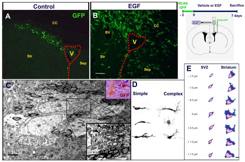
Figure 3. SVZ astrocytic stem cells give rise to EIPs.
GFP-expressing cells after vehicle (A) and EGF (B) infusions. A number of GFP+ cells derived from the SVZ astrocytes were observed around the lateral ventricle (V) and infiltrated the brain parenchyma of EGF-infused animals. C. Axonal tracts showing two GFP-labeled cells analyzed by EM. Upper inset shows the semi-thin section from where the ultrathin section was obtained. Lower inset: Higher magnification of the area indicated in C; arrowheads indicate DAB precipitates. D: Z-stack reconstructions of GFP+ cells. E: Schematic 3-D reconstructions by EM. The numbers at the left indicate the distance between each section. Time line and mouse brain schematic drawing that shows the cannula’s spatial position and the injection site to label SVZ astrocytes CC: Corpus callosum; Str: Striatum; Sep: Septum; BV: Blood vessel. Scale bar in B = 100 μm; C = 2.5 μm, inset = 1 μm; E = 5 μm.

