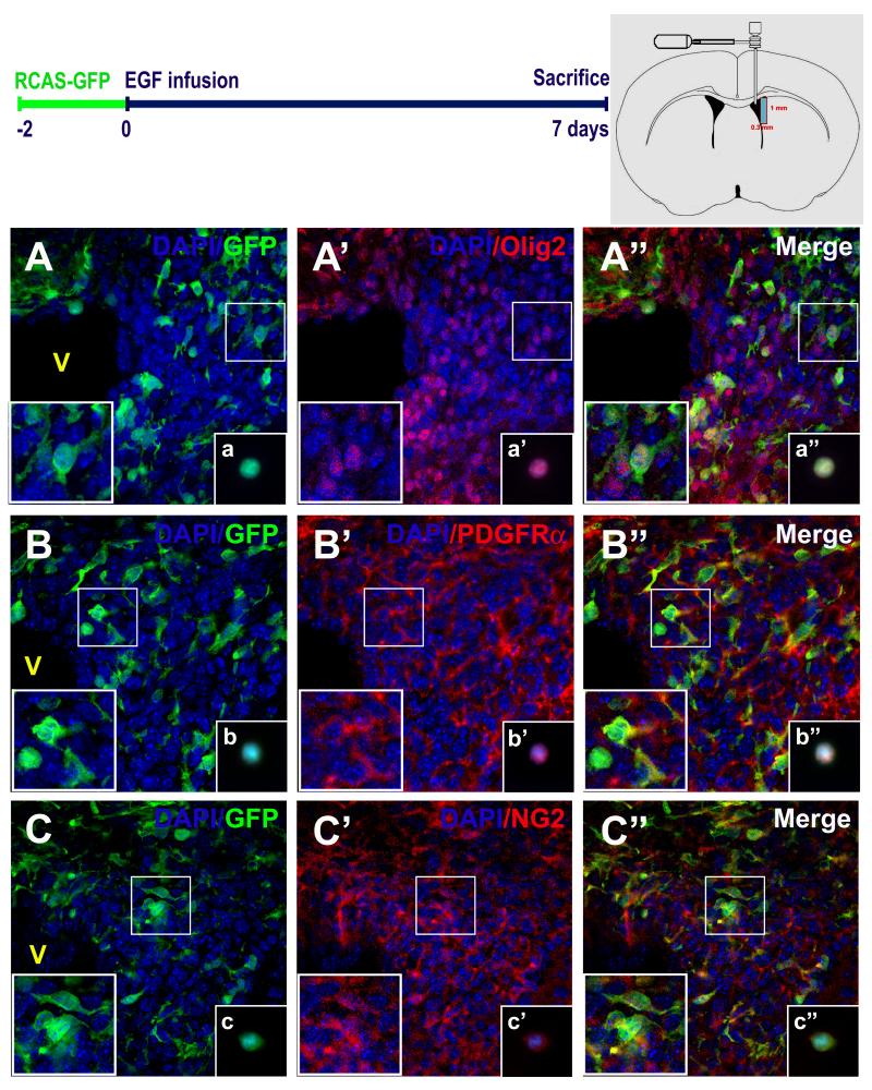
Figure 4. EIPs express markers related to oligodendrocyte lineage.
Brain sections were co-stained with anti-GFP and anti-Olig2 (A - A“), anti-PDGRFα (B - B”), and anti- NG2 (C - C“) antibodies. High magnification cell details are shown at bottom left square. Independent experiments and immunolabelings were performed on freshly-dissociated cells (insets: a-a”, b-b“, c-c”). Schematic brain drawing depicts the dissected area (1mm × 0.3 mm) where fresh-dissociated cells were obtained. Nuclear staining was performed with DAPI (blue). V: Ventricle. Scale bars = 20 μm.

