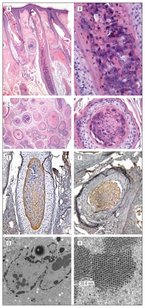
Figure 2.
Histologic features. A–D, Hematoxylin-eosin staining of tissues in vertical (A, low power; B, high power) and horizontal (C, low power; D, high power) sections demonstrates aberrant keratinization of the inner root sheath hair shaft cells, with numerous enlarged, bulbous anagen hairs and a thin layer of basophilic, germinative cells transitioning to inner root sheath–type cells containing several enlarged bluish gray inclusions. Vacuolated keratinocytes with pyknotic nuclei and coarse keratohyaline granules also are seen. E and F, Immunohistochemical staining for the polyomavirus middle T antigen on both vertical (E) and horizontal (F) sections demonstrates strongly positive staining of cellular inclusions within keratinocytes composing the inner root sheath, confirming the presence of polyomavirus. G and H, Scanning electron microscopy reveals small (35.6-nm), icosahedral, regularly spaced, intracellular viral particles within these inclusions, consistent with polyomavirus.

