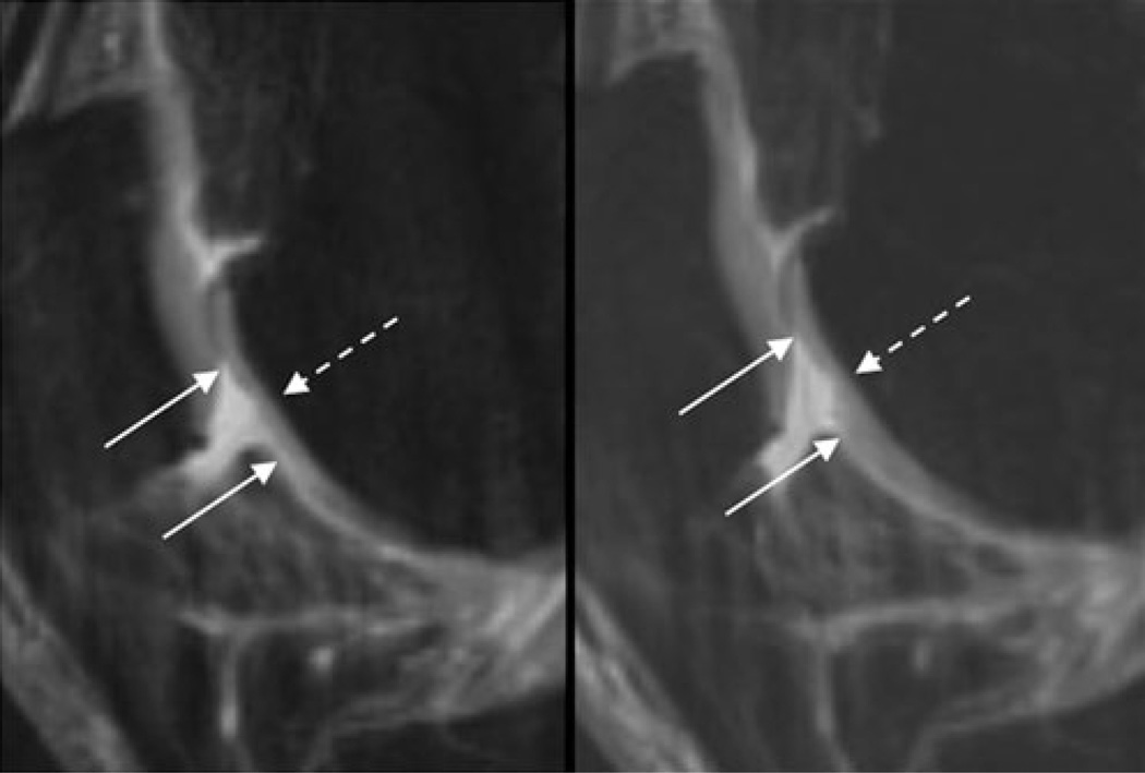
Fig. 2.
Sagittal T2w-FSE images (TR/TE 3,600/8.5 ms) of the right trochlea in a 50-year-old female OA patient at BL (left) and after 12 months (FU1, right). At both time points, a 12-mm-wide partial thickness defect of the cartilage is shown (the maximal extent of this lesion in cranio-caudal extent is outlined by the solid arrows); therefore, a WORMS of 3 has been assigned. At BL, only the superficial layer of the cartilage is affected, whereas at FU1 the middle and parts of the basal layer are also impaired, resulting in a semi-quantitative estimated lesion size of 53 mm3 at BL and 126 mm3 at FU1

