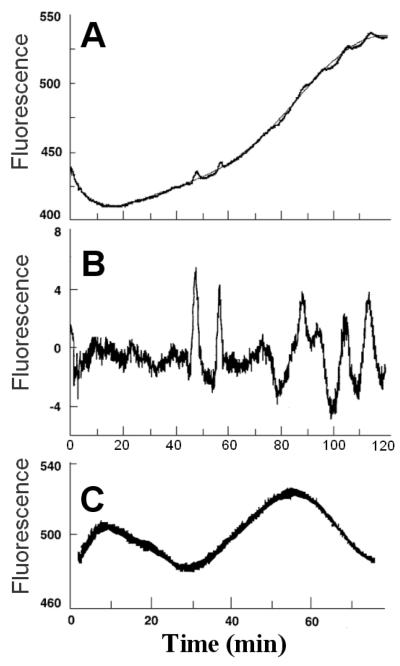
Figure. 1. Oscillations in NADH fluorescence in a cell-free supernatant prepared from isolated rat islets.
A and B are traces obtained from the same extract, and C represents a separate extract. The raw data in A were fitted with a 6th order polynomial (narrow line), which was subtracted out in B to show the repetitive oscillations more clearly. Data are arbitrary fluorescence units. The reaction mixture contained 1 mmol/l ATP, 1 mmol/l MgCl2, 20 mmol/l HEPES, pH 7.1, 100 mmol/l KCl, 7.5 mmol/l potassium phosphate, 10 mmol/l glucose, 0.5 mmol/l NAD, 0.06 U/ml crystalline yeast hexokinase (gel filtered in 20 mmol/l HEPES), 0.04 U/ml apyrase (an ATPase), and islet extract equivalent to 50% of the volume. In C, the HEPES buffer was pH 7.2. Reactions were started by adding hexokinase, apyrase, and islet extract in rapid succession.

