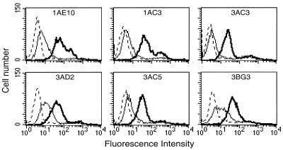
Figure 3.
TH1 Vβ8+CD4+ clones and TH2 Vβ8+CD4+ clones express similar levels of Qa-1 on their surface. TH1 (Upper) and TH2 (Lower). CD4+ T cell clones were stained with antisera to Qa-1a (bold solid lines), Qa-1b (thin solid lines), or with normal B10.PL serum (dotted lines) as described in Materials and Methods. CD4+ T cell lines were stained on day 6 after activation.

