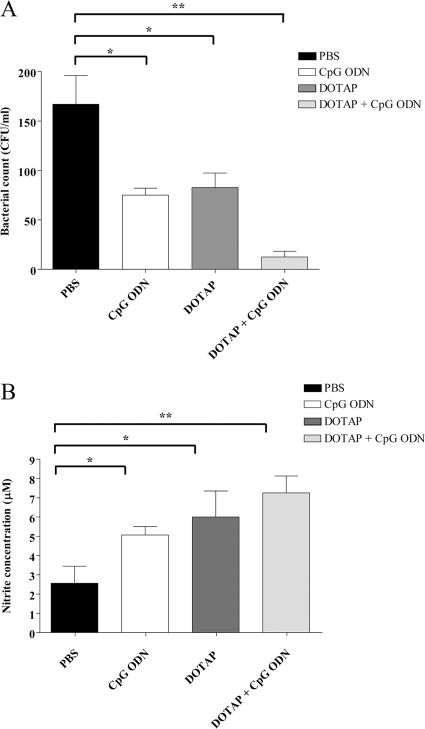
Fig 5.
Intracellular bacterial survival and nitric oxide production of macrophages. Numbers of intracellular bacteria (A) or nitrite concentration (B) of splenic macrophages from mice treated with PBS or CpG ODN on day −2 or with DOTAP alone or ODN plus DOTAP on day −30 is shown. Macrophages (1 × 10⁶ cells/ml) were infected with B. pseudomallei at an MOI of 10:1 for 1 h. Extracellular bacteria were removed by incubation with medium containing 250 μg/ml kanamycin for 2 h. The medium was then replaced with new medium containing 20 μg/ml kanamycin and further incubated for 5 h. At 8 h, the supernatants were assayed for nitrite, and macrophages were lysed with 0.1%Triton X-100 and cultured on nutrient agar to determine numbers of intracellular bacteria. The levels of nitrite were determined using the Griess reagent. Each bar represents means ± SD. Results from 3 independent experiments are shown. The asterisks (* and **) indicated significant differences from results for the PBS control (P < 0.05 and P < 0.01, respectively).

