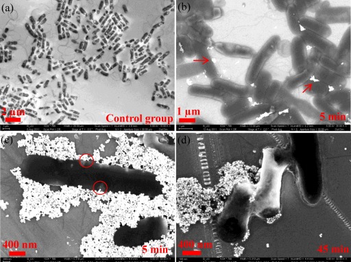
Fig 1.
SEM images of E. coli cells with and without hematite treatment at different exposure times (indicated in the bottom right of each legend). (a) Untreated E. coli cells. (b) E. coli cells with light attachment of hematite NPs (white dots, as indicated by the arrows). (c) E. coli cells with heavy sorption of hematite NPs. (d) Deformed E. coli cells.

