Figure 1.
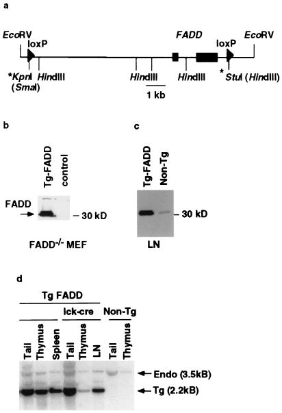
Generation of T cell-specific FADD−/− mice. (a) A schematic diagram of the 12-kb EcoRV genomic fragment from the mouse FADD locus used to reconstitute FADD−/− mice. The two rectangular boxes represent the two exons that encode the FADD protein. Arrowheads represent the loxP sites. The * at the 5′-end denotes the loxP and a SmaI site introduced at the KpnI site, and the * at the 3′-end denotes the loxP and a HindIII site introduced at the StuI site. (b) Western blot analysis of FADD expression in FADD−/− MEF cells. FADD-deficient MEF cells were transiently transfected with the 12-kb FADD-loxP construct or an irrelevant DNA control. Whole cell extracts were made 2 days post-transfection. FADD expression was assessed by a Western blot analysis with anti-mouse FADD rabbit antibodies. Arrow indicates the phosphorylated and unphosphorylated FADD proteins. (c) Whole cell extracts from lymph nodes (LN) of line 33 FADD transgenic mice or a nontransgenic FADD+/− littermate control were subjected to Western blot analysis by using anti-FADD rabbit polyclonal antibodies. (d) Southern blot analysis of genomic DNA from tail, thymus, and peripheral immune organs (either spleen or purified lymph node T cells = LN) of tFADD−/− mice and their transgenic FADD wild-type littermate or nontransgenic FADD heterozygous mice. The genomic DNA was digested with EcoRV and HindIII. The resulting Southern blot was probed with a fragment from the second exon of FADD. Arrows indicate the endogenous 3.5-kb band and the 2.2-kb transgenic band.

