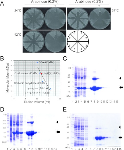
Fig 2.
Characterization of MazE-MazF fusion protein. (A) Toxicity of MazE(42)F-FXa in E. coli. The transformants harboring pBAD24 vector (1 and 8), mazF-ec (2 and 7), mazF-bs (4 and 5), and mazE(42)F-FXa (3 and 6) were streaked on M9 plates with or without 0.2% arabinose and incubated at three different temperatures, 24, 37, and 42°C. (B) Gel filtration of MazE(42)F-FXa. The linear trend line is used for calculating the molecular mass for the MazE-MazF fusion protein. The purification of MazE(42)F-FXa (C), MazEF-HIV (D), and MazEF-HCV (E) are also shown. Lane 1, molecular mass markers; lane 2, the whole-cell lysate without induction; lane 3, the whole-cell lysate after incubation for 4 h at 37°C; lane 4, the cell pellet; lane 5, flowthrough fraction; lane 6, wash fraction; and lanes 7 to 15, elution fractions. Arrows indicate the positions of monomers of the fusion proteins, and arrowheads indicate the positions of their dimers.

