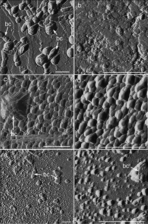
Fig 3.
AFM deflection images (taken in air) of bacterial calcite precipitates formed on calcite single crystals. (a) Calcified B. diminuta cells (calcified bacterial cells [bc]); (b) detail of oriented calcite overgrowth formed in the presence of B. diminuta; (c) precipitates (growth islands) formed in the presence of the bacterial community, including a micrometer-sized growth structure associated with a calcified bacterial cell; (d) detail of oriented calcite growth islands formed in the presence of the bacterial community; (e) precipitates formed in the presence of M. xanthus, including micrometer-sized calcified bacterial cells and 2D nuclei; (f) detail of 2D nuclei (growth islands) formed in the presence of M. xanthus. Bars, 2 μm.

