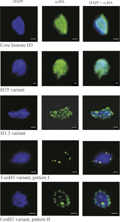
Fig 2.
Localization of T. vaginalis core H3-HA and H3-HA variants in interphase nuclei. T. vaginalis mid-logarithmic-growth population was used for detection of tagged proteins. All cells displayed homogeneous labeling of all interphase nuclei in which core H3-HA (TVAG_270080) and H3T-HA (TVAG_185390) were expressed. The same punctate pattern was observed throughout the population that expressed H3.3-HA (TVAG_087830). Images are representative of tagged protein localization observed in over 200 cells per each strain; localization demonstrated in the figures was observed in 90% of examined cells. Two distinct patterns were observed for trichomonads that expressed CenH3 (TVAG_224460); patterns I and II were observed in 20% and 80% of examined cells, respectively. Bars, 1 μm.

