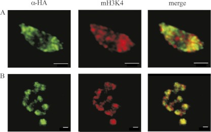
Fig 4.
Active regions of transcription visualized by monomethyl H3K4 immunostaining and localization of the T. vaginalis H3.3 variant (TVAG_087830). (A) Interphase nuclei; (B) metaphase chromosomes. Images are representative of tagged protein localizations observed in over 200 cells per each strain. Bars, 1 μm.

